A patient in the ICU reports worsening shortness of breath. Does this subxiphoid view echocardiogram suggest cardiac tamponade as the etiology of the complaint?
Tamponade? from MDT on Vimeo.
#Cardiac tamponade physiology on echocardiogram can be difficult to distinguish from just a moderate to large pericardial effusion
#This is all about ultrasound, so forget about those ‘clinical signs’ like hypotension, distant heart sounds, pulsus paradoxus, and visible JVD. A sonographic finding with strong specificity for cardiac tamponade is right ventricular wall collapse during DIASTOLE
#Check out this slightly dizzying image showing how M-mode can be used to draw a line through the RV free wall, septal wall, and mitral valve to record their motion in real time
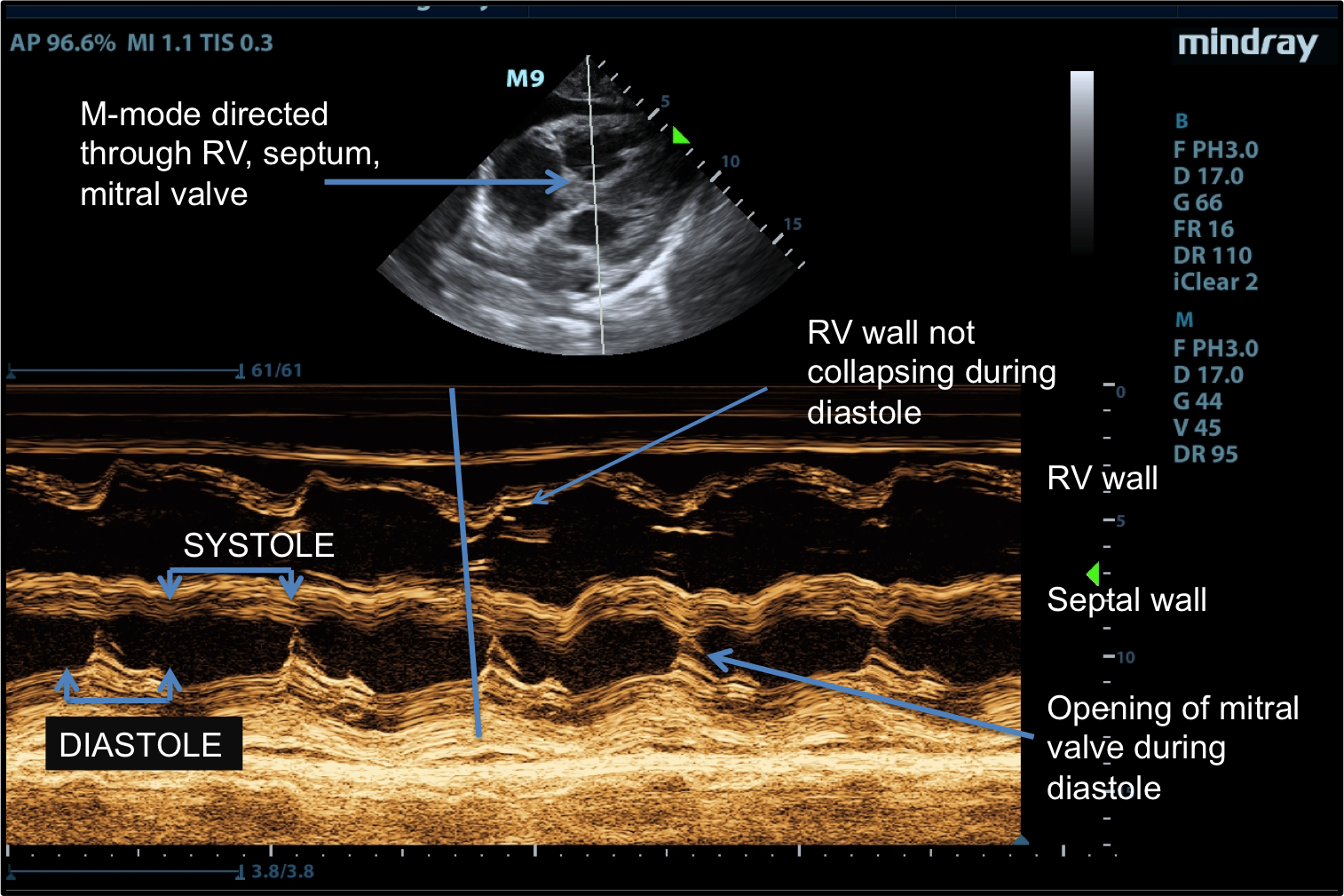
#Remember that Diastole is when the mitral valve is OPENING toward the septum
#The RV free wall collapses appropriately during the END of Systole, which is the moment of maximum endocardial contraction
#The RV wall then expands outward during Diastole. In tamponade, this wall would collapse during Diastole due to the high pressure of a pericardial effusion. Therefore this echocardiogram does NOT show tamponade (womp womp, major letdown, but certainly better for the patient). Beware of patients with a known history of pulmonary hypertension and/or RV failure as they will likely have thick RV walls, which will complicate the sonographic evaluation.
References
![]()